Active movement of the trunk. The delicate spinal cord is housed within the spinal canal of the bony spine and provides the nerve connections throughout the body.
 Pin On Occupational Therapy
23012018 The vertebral column also known as the spinal column is a flexible column that encloses the spinal cord and also supports the head.

Genuine spine diagram and the description. The top part of the spine has seven vertebrae C1 to C7. The vertebral column is divided into 5 different regions - cervical thoracic lumbar sacrum and coccyx. 31052021 Spinal cord diagram The spinal cord is a continuation of the brainstemIt extends from the foramen magnum at the base of the skull to the L1L2 vertebra where it terminates as the conus medullaris medullary cone.
The spines four sections from top to bottom are the cervical neck thoracic abdomen lumbar lower. These neck vertebrae allow you to turn tilt and nod your head. The spinal cord is a bundle of spinal nerves wrapped together.
Several spinal nerves emerge out of each segment of the spinal. The illustration is available for download in high resolution quality up to 5000x5000 and in EPS file format. The 33 vertebrae make up five distinct spine segments.
Download this image now with a free trial. It contains the osteology arthrology and myology of the spine and back. The spinal nerves enter and exit the spinal cord through small spaces between the vertebrae.
It is particularly interesting for physiotherapists osteopaths rheumatologists neurosurgeons orthopedic. The spine serves the important functions of. The 10 spinal laminae of the spinal cord are shown on a second diagram about the grey matter of the spinal.
Lumbar Spine Anatomy Video. Can be used for personal and commercial purposes according to the conditions of the purchased Royalty-free license. Human Spine in front diagram with the name and description of all sections of the vertebrae and segments vector illustration.
Diagram of a human spine with the name and description of all sections of the vertebrae. Human Spine in front diagram with the name and description of all sections of the vertebrae and segments vector illustration. Royalty-free stock vector ID.
Spine - Spinal Stenosis Detailed anatomy of the human brain. This is an editable EPS 10 vector illustration with CMYK color space. The blood vessels which carry oxygen to the spinal cord also use these spaces.
22092020 The anatomy of the grey matter of the spinal cord is summarized on a diagram with the various grey matter nuclei note that this representation is virtual because some nuclei are only present at some levels between them but are displayed on a single cross-sectional diagram. The size and shape of each lumbar vertebra is designed to carry most of the bodys weight. It forms a vital link between the brain and the body.
A thin thread called filum terminale extends from the tip of the conus medullaris all the way to the 1st coccygeal vertebra Co1 and anchors the spinal cord in. Each structural element of a lumbar vertebra is bigger wider and broader than similar components in the cervical and thoracic regions. You have 8 pairs.
22092020 This human anatomy module is composed of diagrams illustrations and 3D views of the back cervical thoracic and lumbar spinal areas as well as the various vertebrae. Understanding the anatomy of your lower spine can help you communicate more effectively with the medical professionals who treat your lower back pain. More similar stock illustrations Human nervous system medical vector illustration diagram with parasympathetic and sympathetic nerves and connected inner organs.
The cervical spine makes an inward C-shape called a lordotic curve. Starting at the neck and going down toward your buttocks rear end these segments include. Vector image Diagram of a human spine with the name and description of all sections of the vertebrae.
Spinal cord diagram stock illustrations. 10022015 Together the brain and spinal cord make up the central nervous system. By definition the vertebrae are the bones of the spine or backbone that function to protect the spinal cord and support the rest of the body and cranium.
In adults the spinal cord is usually 40cm long and 2cm wide. Spine Diagram A diagram of a human female spine showing a side view of the vertebra of the spinal cord within the the body. The lumbar region of the spine more commonly known as the lower back is situated between the thoracic or chest region of the spine and the sacrum.
29042020 The spine also known as the vertebral column or spinal column is a column of 26 bones in an adult body 24 separate vertebrae interspaced with cartilage and then additionally the sacrum and coccyx. The spinal cord is divided into five different parts. The spine has ligaments that limit motion and muscles that attach to provide motion.
Plus get full access to a library of over 316 million images. 07122020 The vertebral column also known as the spinal column is made up of 33 vertebrae. The Lumbar Spine has 5 vertebrae abbreviated L1 through L5 largest.
 Pin On Spine
 Pin On Thoracic Vertebrae
 Pin On Medical Illustration
 Pin On Body Structure
 Pin On Reflexology
 Cervical Vertebra C1 C7 Anterior View Cervical Vertebrae Vertebrae Skull And Bones
 Pin On Ministry Helps
 Pin On Body Anatomy Massage
 Pin On Medical Health
 Pin On Axis
Pin On Ot Spinal Cord Injury
 Pin On Vertebral Column
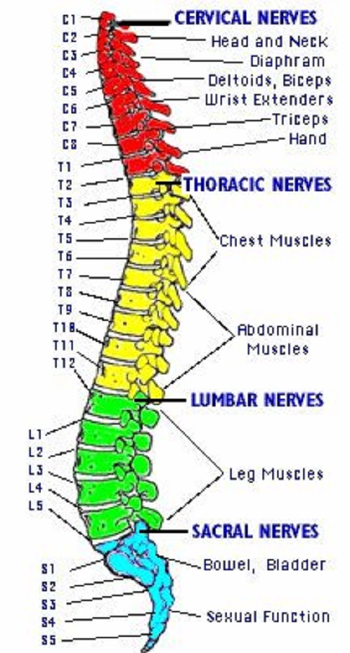 Diagram Bone Diagram Back Full Version Hd Quality Diagram Back Diagrammi Poliarcheo It
 Pin On Lumbar Vertebrae